Radiographers use this interface to take X-ray images. The previous version was badly overloaded: every new feature had been bolted on top of the old one without rethinking the flow. Because of that, medical staff had to work through a multi-page manual before they could start.
NDATHE LOGO AND PATIENT DATA ARE COVERED. EVERYTHING ELSE IS SHOWN AS IT IS.
01How I worked it out
I ran interviews in hospitals and watched the staff work on site. I looked for where people stumbled, which features they never used, and which ones they did not even know existed. Separately I collected the positive feedback — what worked and what a redesign must not break.
01Interviews with hospital staff
02Observing real work on site
03A/B testing of the solutions
04Customer journey map (CJM)
02What came out of it
Part of the functionality went unused not because nobody needed it, but because nobody knew about it: it existed only inside the multi-page manual. The interface offered no onboarding, was hard to make sense of, and was visually overloaded on top of that.
01Features people did not know existed
02Points where the work stalls
03Features deliberately left unused
The study journal
BEFOREAFTER
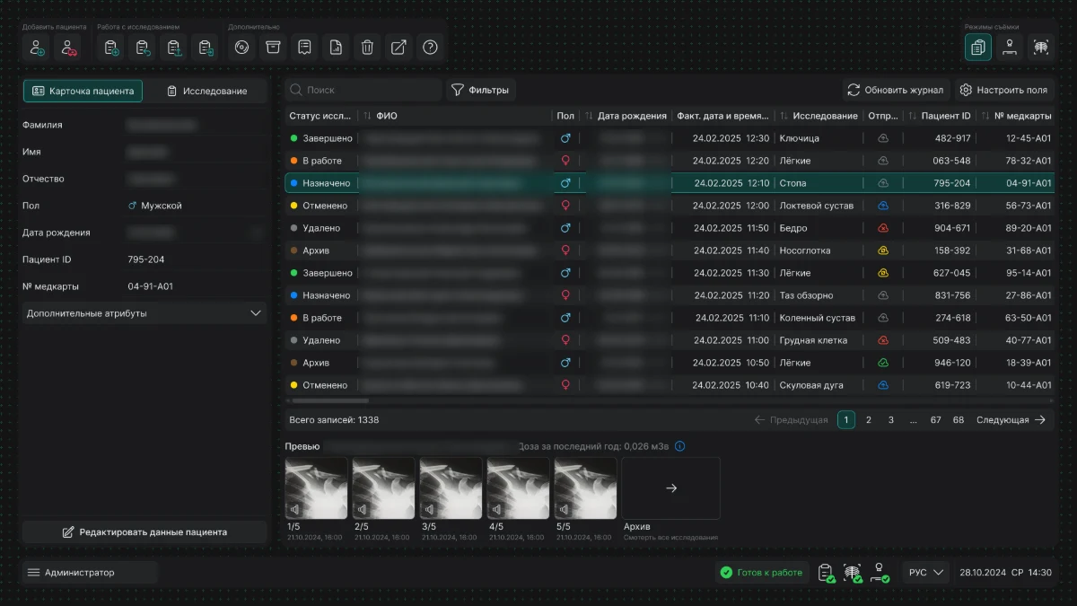
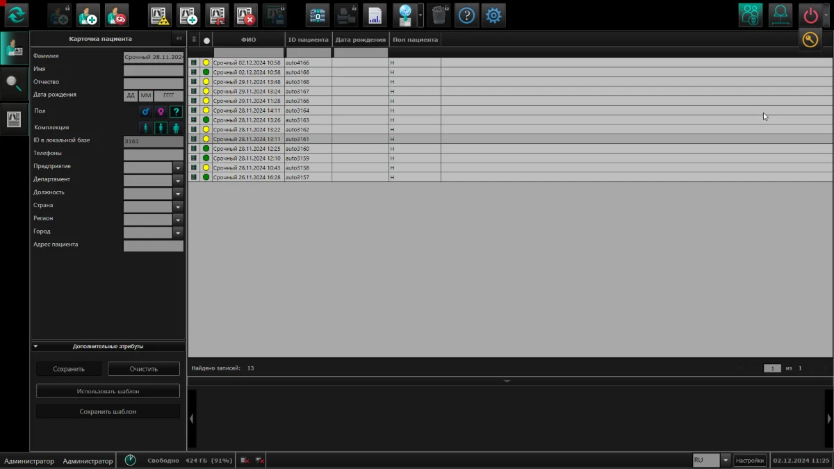
The toolbar is split into labelled groups, and the flow now reads
The patient card got shorter and the table easier to scan
Image previews appeared, along with the accumulated annual radiation dose
03The redesign
Together with the second designer we put together a new version of the screens: stripped away the layers of old and new flows, gave the useful features their visibility back, and removed what was only taking up space. We got the first screens approved by the stakeholder and worked through the rest from there.
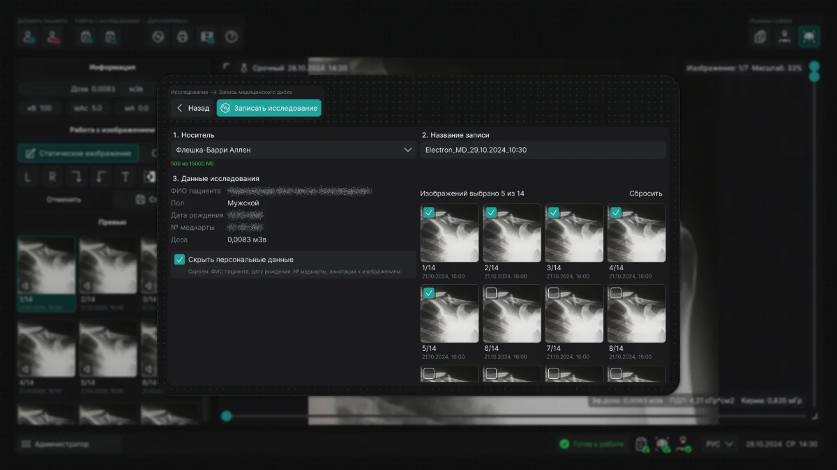
Burning a study to disc: the medium, the record name, the choice of images
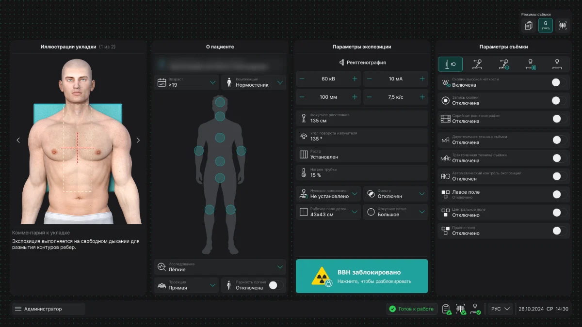
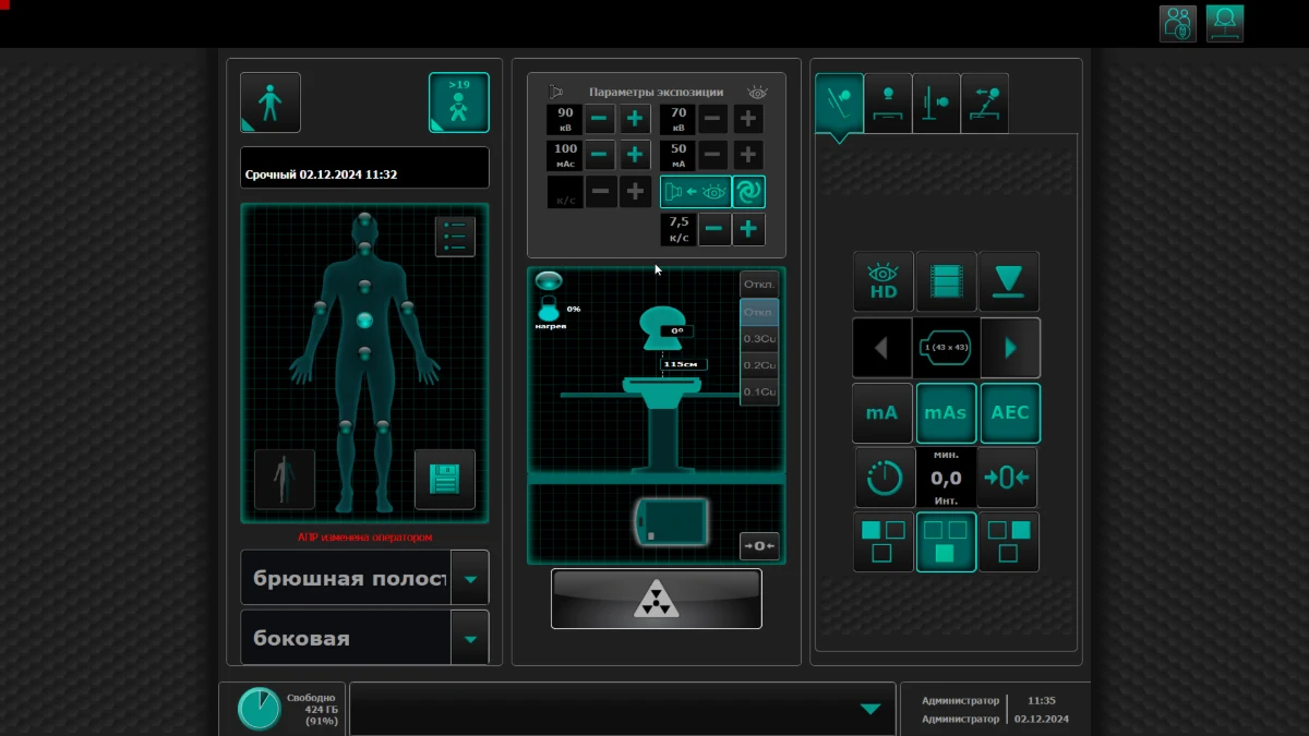
The imaging parameters screen
BEFOREAFTER
The screen is split into four labelled panels
A positioning illustration with a comment appeared
Imaging parameters can be read at a glance
The high-voltage button is highlighted and labelled
04The link to the system
It was this redesign that made it obvious the screens would drift apart again without a shared library. The UI kit grew out of these screens and later became a design system — that is a separate case, 01. The radiographer interface is already assembled on it.
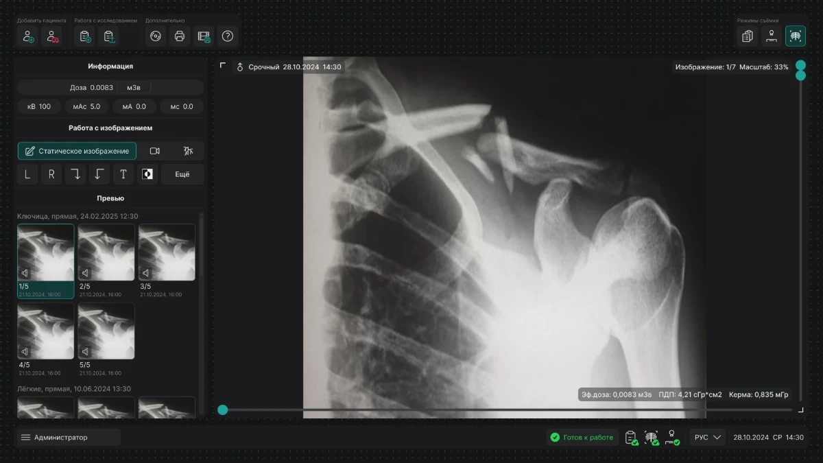
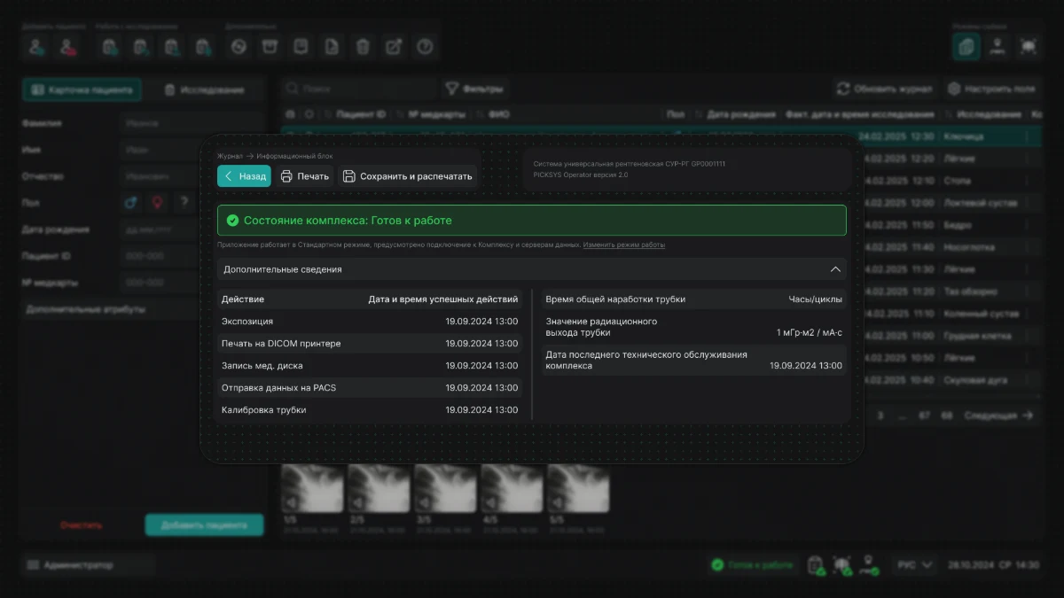
Viewing an image: measurement tools on the left, dose and parameters under the pictureThe info panel: system status, tube usage, last service date
RESULT
Redesign on our own system
The interface became better looking and more modern, and after A/B testing I confirmed that people understand it better than the old version.
Anastasia Kuratnik
Product Designer (UX/UI)
Four years in design, a year and a half of them in product. I design complex desktop and web interfaces, build design systems and get them adopted. I run research: in-depth interviews, surveys and usability testing — with doctors and domain specialists. I have worked with clients from Belarus and Kazakhstan.
LOCATION
Saint Petersburg, open to relocation
FORMAT
Remote, hybrid or on-site
EMPLOYMENT
Full-time
LANGUAGES
Russian — native, English — intermediate
EXPERIENCE
«Electron»
May 2025 — present
Product designer, design system designer · Medical diagnostic equipment · Saint Petersburg
I own the interfaces of X-ray equipment at every stage: researching how doctors actually work, design, handover to engineering, design review.
The devices in the product line had grown apart from each other — there was no shared component library at all.
Made the case to stakeholders for moving to a single design system, with an estimate of time and development cost. Got approval and started the project.
Built the design system and wrote the technical documentation for engineering.
Cut the time a designer needs to assemble a typical screen feature from a week down to three working days.
Used it as the basis for redesigning the interfaces across the company’s whole product line.
Ran in-depth interviews and surveys with doctors and domain specialists — and corrected the user flows before development started.
Produced wireframes and interactive prototypes within the constraints of the desktop platform.
Pyrus agency
February 2025 — February 2026
UX/UI designer · Marketing agency · Belarus
Landing pages, multi-page sites and promo pages with responsive versions; banners, social media, ad creatives, presentations, outdoor advertising.
Ran competitor and audience analysis, and built user flows on top of it before moving to layouts.
Designed logos and identity, worked with clients’ brand books, prepared micro-animations and handed layouts over to development.